Your veterinarian will perform three tests to determine whether or not your dog has a ruptured or torn cruciate ligament.
Tests to diagnose ruptured or torn cruciate ligament include:
- Physical exam
- X-rays
- Manual instability tests.
Physical Exam
Your vet will look for the following during the physical exam:
- Positive Sit Test: Dogs normally sit with the stifle fully flexed under the pelvis. Dogs with torn cruciate ligament injury will frequently sit with the entire leg out to the side. Sitting with “the entire leg out” is referred to as a “positive sit” test.
- Medial Buttress: By palpating the inside of your dog’s stifle, your veterinarian will be able to discern the presence of a medial buttress. A medial buttress is a swelling on the inside of the stifle developing in an attempt to control the instability. Swellings appear as early as one month after cruciate injury.
- Stifle swelling called effusion: Swelling inside a joint is termed effusion. Effusion develops as a reaction to the torn fibers. It presents as a swollen knee. Your veterinarian will be able to easily discern the presence of effusion during the physical exam. When effusion exists, damage to the cruciate ligament is confirmed.
X-rays
Your veterinarian will most likely take x-rays to positively diagnose cruciate disease.
X-rays of the stifle joint can determine the presence of joint effusion or arthritis.
Both disorders are diagnostic of cruciate damage.
Manual Testing for Instability
The first test is a cranial drawer.
During the cranial drawer test, your vet will stabilize your dog’s femur with one hand and at the same time manipulate your dog’s tibia with the other hand.
If your dog’s tibia moves forward, the result is a positive drawer.
When the torn cruciate ligament is ruptured, the tibia moves similar to a drawer being opened.
If the rupture happened a while ago, the side of the leg facing the opposite leg will be swollen.
Swelling on a leg facing the opposite leg indicates arthritis and is called medial buttress.
The majority of dogs with cranial drawer signs can be palpated and examined with the dog awake.
However, if any uncertainty exists, palpation of the dog under sedation or anesthesia is necessary.
Dogs with partial tears of the cranial cruciate ligament and dogs with chronic cranial cruciate ligament injury will tend to have less cranial drawer than the “normal” dog with a cranial cruciate injury.
It is imperative to compare the drawer of one stifle to the opposite stifle. The second manual test is termed cranial tibial thrust.
The cranial tibial thrust test is another manually applied force to test the stability or lack of stability of the stifle joint.
So, all of the above tests should give your veterinarian a good idea of whether or not a ligament injury is causing your dog’s lameness.
Due to the progressive nature of ligament injury, dogs present to the veterinarian with either a partial or complete tear.
Diagnosing a Partial Tear
A partial tear occurs when individual fibers of the ligament tear, not the entire ligament. Imagine your daughter’s ponytail.
The entire ponytail represents the entire cranial cruciate ligament, and every single hair in the ponytail represents an individual fiber in the cruciate ligament.
When individual fibers tear and lameness occurs, the situation is described as a partial tear.
Partial tears lead to lameness and swelling but rarely lead to gross instability of the joint.
Some dogs adapt to partial tears and no surgery is necessary.
Diagnosing a Full Tear or Rupture
Occasionally, a full tear is the result of trauma much like the tear seen in football players.
However, as mentioned earlier, partial tears frequently escalate to full tears.
Full tears lead directly to joint instability and generally require costly surgery to correct.
The cost can vary depending on the surgeon and the surgical technique employed.
The treatment for each type of tear will depend on a variety of factors including the size and weight of your dog, your dog’s age, other concurrent diseases, your veterinarian’s experience, and more.
Diagnosing a cranial cruciate ligament injury usually requires fairly straightforward tests such as a complete physical exam and x-rays.
Unfortunately, the treatment may be much more complex.
Proactive Ways to Help Your Dog’s Ligament Challenges
There are many quick and easy changes you can make at home to help you give your dog an edge on easing tendon and ligament challenges.
- Learn more about torn ligaments and cruciate disease.
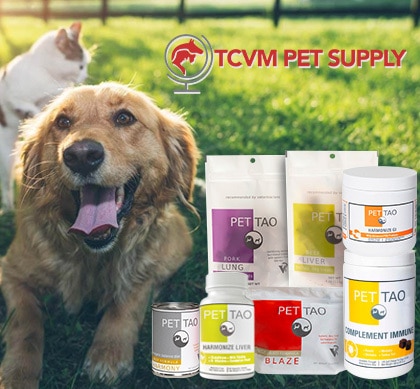
- Provide joint support. PET | TAO’s Harmonize Joint is a blend of Eastern herbs and Western supplements working together to lubricate and restore your dog’s joints.
- Ease your dog’s discomfort naturally. PET | TAO’s Comfort is a blend of Eastern herbs and Western supplements to soothe your dog’s arthritic challenges to make him/her more comfortable.
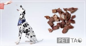
- Try PET | TAO Freeze Dried Beef Liver Treats. According to TCVM, the liver controls tendons and ligaments. As few as 5-6 treats per day can make a huge difference in your dog’s tendon and ligament health!
- Try a Blood-building TCVM Diet. PET | TAO Zing dog food builds Blood. According to TCVM, Blood deficiency leads to ligament tears.
- Learn more about TCVM Herbal Remedies. Chinese medicine offers many amazing natural solutions for ligament and cruciate challenges. Some good examples are: